What do visual representations of clubbing tell us about a disorder? Visual aids of this condition can be essential tools in diagnosis and education.
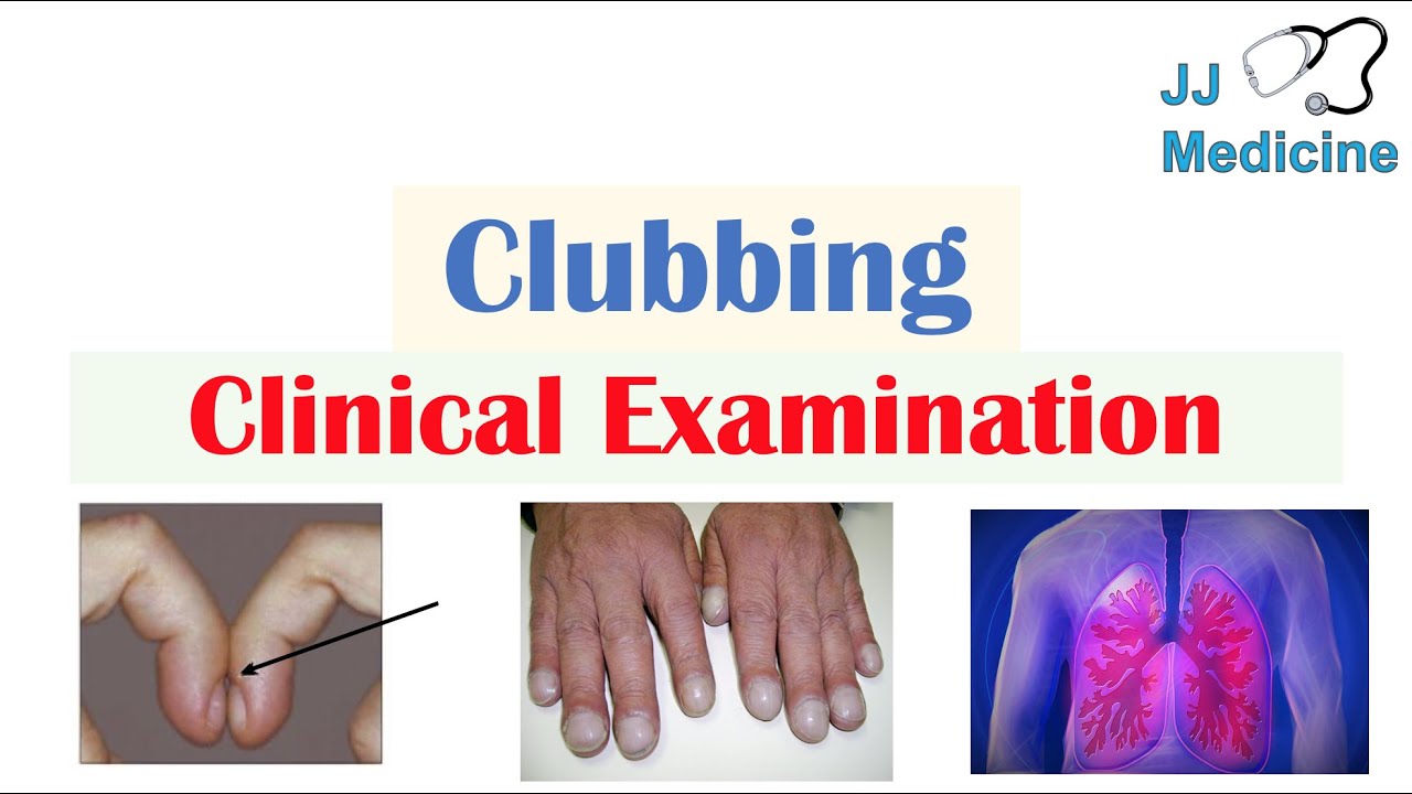
Images depicting clubbinga condition characterized by the enlargement of the fingertips and toescan be valuable diagnostic aids. These images, whether photographs or medical illustrations, document the physical manifestation of the disorder. They allow healthcare professionals to compare observed changes with established visual markers of clubbing. Examples might include images showcasing the characteristic bulbous shape of the distal phalanges or the angle formed at the nail base. These visual cues can facilitate a quicker and more accurate diagnosis by supporting clinical findings.
The utility of these images extends beyond diagnosis. Educational resources utilizing clubbing disease pictures can be crucial in raising awareness about the condition among patients and healthcare providers. Visual representations can convey the essence of the condition more effectively than written descriptions. Images can be used in medical textbooks, patient handouts, and online resources. Showing various stages of the condition's progression could highlight the importance of early intervention and appropriate treatment. Historical context reveals the importance of visual documentation. Medical illustration, particularly, has played a key role in recording and disseminating knowledge about clubbing throughout history.
Moving forward, analysis of different types of clubbing and the related clinical symptoms can be enhanced with this imagery. These resources can support clinical training and research initiatives, by allowing the demonstration of varying severity and manifestations.
Clubbing Disease Pictures
Visual documentation of clubbing disease plays a critical role in diagnosis, education, and research. Accurate depiction of the condition is essential for various purposes.
- Diagnosis
- Education
- Research
- Documentation
- Progression
- Severity
- Variation
- Comparison
Visual representations of clubbing, through photographs and illustrations, allow for precise documentation of the condition's progression and variation in severity. Comparison of images facilitates diagnosis, assisting healthcare professionals in identifying subtle changes in affected tissues and comparing presentations across different cases. Educational materials incorporating such pictures aid in raising awareness and informing patients about the disease, clarifying diagnostic criteria, and facilitating better patient understanding. Images also support research by providing standardized visual data for analysis, enabling tracking of disease progression and identifying correlations with various clinical factors. Illustrations of clubbing can thus serve as essential tools in the diagnosis, education, and study of this particular medical condition.
1. Diagnosis
Accurate diagnosis of clubbing relies heavily on visual assessments. Images of clubbed fingers and toes are integral components in this process, supplementing clinical examination and aiding in differentiation from other conditions presenting with similar physical manifestations. A thorough understanding of these visual features is critical for proper diagnosis and subsequent management.
- Visual Identification of Clubbing:
Visual representations of clubbing allow for objective comparison of observed changes in the affected digits with established visual markers. Photographs and illustrations showcase the characteristic enlargement of the distal phalanges, the changes in the angle at the nail base, and the shape of the nails. These visual cues facilitate a more rapid and accurate diagnosis, complementing traditional clinical evaluations. Images of healthy controls and those with various stages of clubbing are essential for differentiating normal variations from the specific changes seen in this condition.
- Differential Diagnosis:
Visual comparison with images of other conditions exhibiting similar physical features is critical for differential diagnosis. Clubbing presents uniquely in imagery, and these images help clinicians rule out other possible causes. Comparison with images of conditions that can mimic clubbing, like certain inflammatory diseases, helps refine the diagnostic process. Accurate visual representations assist in the elimination of incorrect diagnoses and reduce the potential for misinterpretation.
- Progression Monitoring:
Images documenting the progression of clubbing over time can offer crucial insights for both diagnosis and treatment. Monitoring changes in the affected digits through repeated visual documentation can provide evidence of disease progression. Observing the evolution of clubbing via visual representations can aid in refining the approach to treatment, particularly in identifying treatment efficacy and adjusting management strategies over time.
- Educational Value:
Visual representations of clubbing serve as invaluable educational tools for healthcare professionals. Images in medical textbooks, clinical training materials, and online resources aid in building a shared understanding and recognition of the specific morphological changes associated with clubbing. This can enhance diagnostic accuracy across various settings and improve patient care by fostering accurate interpretation and consistent application of diagnostic criteria.
In conclusion, the inclusion of "clubitis disease pictures" as a critical element of the diagnostic process enhances accuracy, facilitates differential diagnosis, and enables continuous monitoring of disease progression. These visual aids play a vital role in ensuring appropriate and timely intervention, contributing significantly to improved patient outcomes.
2. Education
Effective education concerning clubbing relies on clear and accessible information. Visual aids, such as images of clubbing, play a critical role in this educational process. Understanding the visual characteristics of the condition is crucial for both healthcare professionals and patients.
- Visual Recognition of Clubbing:
Images allow for the identification of key morphological changes associated with clubbing. Visual learning facilitates rapid recognition, enhancing diagnostic capabilities among healthcare professionals. Examples include illustrations highlighting the bulbous distal phalanges, the altered angle at the nail base, and characteristic nail bed changes. Consistent visual exposure to these features improves the ability to discern clubbing from normal variations and other conditions with overlapping physical characteristics. This, in turn, leads to earlier and more accurate diagnoses.
- Differentiating Features:
Educational resources featuring diverse images of clubbing, including varying degrees of severity, are valuable in differentiating the condition from similar-appearing conditions. Comparison between images of clubbing and images of non-clubbing conditions highlight distinctive physical markers. This approach clarifies the subtleties distinguishing the particular morphological characteristics of clubbing from those of related pathologies. Such comparisons, illustrated visually, enable more precise diagnostic decision-making.
- Understanding Progression:
Images documenting the development of clubbing over time provide insights into the condition's progression and impact. Educational resources using such illustrations demonstrate how the condition may evolve over time. Examples showing sequential stages of clubbing help healthcare professionals and patients better understand the potential trajectory of the condition. This dynamic approach strengthens awareness of potential implications and prompts appropriate interventions.
- Patient Education:
Images empower patients by allowing them to visually understand their condition. Patients educated with images of clubbing can actively participate in their healthcare decisions. The ability to interpret relevant medical images fosters a deeper connection with the condition. Visual aids help patients understand the nature of clubbing, enabling them to effectively communicate with their healthcare team and make informed decisions about their management. Illustrations of diverse cases can also show the variety of outcomes and experiences possible for individuals affected by this condition.
In conclusion, "clubitis disease pictures" are not simply illustrative tools; they are integral components of comprehensive education about the condition. The visual nature of the information promotes a robust understanding of clubbing's clinical presentation, facilitating more informed diagnoses, empowering patients, and strengthening overall healthcare strategies.
3. Research
Visual documentation of clubbing, often through photographs and illustrations, is critical to research efforts. Accurate and standardized depictions facilitate comparisons across studies, enabling researchers to identify patterns and correlations. High-quality images aid in quantifying features, such as the degree of enlargement, nail-bed angle, and digit symmetry. This standardized approach allows for the creation of reliable data sets enabling systematic analysis of clubbing and associated factors. Researchers can then draw conclusions about potential causes and mechanisms of the condition.
Furthermore, visual records of clubbing are instrumental in understanding the diverse presentations of the condition. Images of different stages of clubbing, including varying degrees of severity and associated symptoms, can help identify factors influencing the progression of clubbing. Such records support the identification of subtypes or specific patterns associated with different etiologies. Analysis of these images, coupled with clinical data, can enhance the development of diagnostic tools and targeted interventions. Examples include the use of digital image analysis tools to objectively measure changes in nail bed angles or digital photographs used in epidemiological studies investigating the prevalence of clubbing in specific populations. Historical collections of images offer a valuable resource for analyzing trends over time, helping researchers track the effects of specific medical treatments or environmental factors on the condition.
In summary, "clubitis disease pictures" are indispensable tools for research. High-quality visual documentation allows for accurate and standardized comparisons, promoting deeper understanding of disease presentation, progression, and associations. This standardized data collection, in turn, enhances research efficacy, facilitates the development of more accurate diagnostic tools, and ultimately leads to advancements in the management and treatment of clubbing. Consequently, this visual approach is crucial to building a robust knowledge base and further enhancing research efforts related to the condition.
4. Documentation
Comprehensive documentation is essential when studying a condition like clubbing. Visual documentation, in the form of "clubitis disease pictures," provides a crucial element for the accurate and consistent representation of the condition's characteristics. Accurate documentation allows for reliable comparison across different cases, enabling researchers to discern patterns, variations, and potential correlations with other factors. This is particularly important for conditions with subtle or variable presentations, as seen in cases of clubbing. Careful documentation aids in establishing a baseline understanding of the disease's morphology, thereby supporting both diagnostic and research endeavors.
The practical significance of meticulous documentation extends beyond research. Consistent and detailed visual records of clubbing are indispensable tools for clinical practice. Repeated images over time allow for monitoring disease progression, assessing the effectiveness of interventions, and aiding in the differential diagnosis of similar conditions. Clinicians can use these visual records to track changes in the affected digits, objectively evaluating the response to treatment and identifying subtle shifts that might be missed in a purely verbal or written record. Archives of such visual data can also serve as valuable educational resources for training future healthcare professionals.
In summary, the relationship between "documentation" and "clubitis disease pictures" is fundamental to understanding and managing clubbing. Accurate and detailed visual records serve as a cornerstone for diagnostic accuracy, research advancement, and improved patient care. The long-term value of such meticulous documentation cannot be overstated; it facilitates a more complete understanding of the condition and informs future clinical and research strategies concerning clubbing.
5. Progression
Visual documentation of disease progression is crucial, particularly for conditions like clubbing, where subtle changes over time are indicative of disease activity and response to treatment. Images of clubbed digits at various stages provide a visual timeline, allowing for objective assessments of how the condition evolves. This documentation is essential for understanding the disease's natural history and identifying potential correlations between progression and underlying causes or treatments.
Tracking changes in the affected tissues through sequential images enables a deeper comprehension of the dynamics of clubbing. For example, a series of photographs showing gradual enlargement of the distal phalanges, increased nail-bed angles, or changes in the overall shape of the digits can illustrate the progression of the condition. This allows researchers to identify key indicators of disease progression, such as the rate of enlargement or the presence of specific morphologic features at different time points. Comparison of these images with clinical data, like blood test results or treatment regimens, can also reveal potential correlations between progression and influencing factors. This type of longitudinal analysis enhances the ability to establish predictive models or to evaluate the efficacy of particular interventions.
Understanding disease progression through visual documentation is critical for both diagnostic and treatment purposes. The ability to objectively track changes in clubbing helps clinicians monitor the effectiveness of interventions, make informed adjustments to treatment plans, and provide patients with realistic expectations about the anticipated trajectory of the disease. Such documentation also facilitates research by providing valuable data for epidemiological studies and the development of improved diagnostic and therapeutic strategies. Ultimately, recognizing the link between visual progression and the underlying disease process offers opportunities to improve patient outcomes and advance medical understanding of clubbing.
6. Severity
Assessing the severity of clubbing is essential for accurate diagnosis and appropriate management. Visual documentation, through "clubitis disease pictures," plays a crucial role in this process. Images provide a standardized means of evaluating the extent of digit enlargement, nail-bed angle changes, and overall morphological alterations. Severity is directly observable in these representations, influencing diagnostic categorization and treatment strategies. Differences in the degree of clubbing, apparent in the images, directly correlate with underlying disease processes. For example, images demonstrating minimal digit enlargement might suggest a milder condition, while those displaying substantial enlargement may indicate a more advanced disease state, necessitating a more aggressive approach.
The importance of "severity" as a component of "clubitis disease pictures" is highlighted by the ability to track changes. Serial images of the same individual can reveal the progression or regression of clubbing, providing valuable insights into the effectiveness of interventions. Images depicting varying degrees of severity within a specific population can assist researchers in identifying correlations between disease severity and associated factors, like the patient's age or specific underlying conditions. Furthermore, detailed visual records of clubbing severity can aid in developing prognostic models and optimizing therapeutic strategies. The severity observed in the images allows for comparisons across cases and provides a concrete framework for evaluating treatment responses.
In conclusion, "clubitis disease pictures" are indispensable for quantifying the severity of clubbing. The visual representations offer a standardized metric for clinical evaluations and research. Understanding the relationship between observed severity in the images and underlying disease processes enables more targeted interventions and improved patient outcomes. This detailed visual documentation is crucial for researchers, clinicians, and patients alike, providing a tangible basis for comprehending the dynamic nature of clubbing and its manifestations.
7. Variation
Clubbing, a condition characterized by enlargement of the fingertips and toes, exhibits notable variation in its presentation. Visual documentation, through "clubitis disease pictures," is crucial for understanding and characterizing this variability. Recognition of diverse manifestations is essential for accurate diagnosis and treatment planning, differentiating clubbing from other conditions with similar physical characteristics.
- Degree of Enlargement:
Images of clubbing reveal variations in the degree of finger and toe enlargement. Visual documentation allows for comparison across different cases, demonstrating how the condition can manifest as mild, moderate, or severe. Understanding these differences is vital for predicting disease progression and tailoring treatment strategies. Illustrations or photographs showcasing varying degrees of enlargement help to establish thresholds for intervention and monitor the response to therapy.
- Nail Bed Angle Changes:
Visual representations highlight variations in the angle of the nail bed. Images showcase the characteristic deviation from the normal nail-bed angle. Different angles, visible in the pictures, can be indicative of diverse disease processes. Careful observation and documentation of these variations allow for a more precise assessment of the underlying cause of clubbing and the potential severity of the condition.
- Associated Symptoms:
Images may capture the visual presentation of clubbing alongside other symptoms, demonstrating how different patients experience the condition differently. Pictures may show variation in the presence of accompanying symptoms, like pain, skin changes, or edema, which may correlate with certain etiologies of clubbing. This allows clinicians to connect visible physical findings with potential underlying medical causes, further refining the diagnostic process. This variation in associated symptoms is crucial for developing tailored approaches to diagnosis and management.
- Variability in Underlying Causes:
The visual documentation encompasses the broad spectrum of underlying conditions that can cause clubbing. Images alongside clinical information can highlight that clubbing may arise from various conditionscardiac, pulmonary, or other systemic diseases. Diverse causes underpin the visual variability observed, which is crucial to keep in mind. Recognition of this variability aids in targeting appropriate investigations for underlying etiologies.
In summary, the variation inherent in clubbing's presentation necessitates comprehensive visual documentation. "Clubitis disease pictures," by capturing the diverse manifestations of the condition, assist in the identification of patterns and correlations. Recognizing the spectrum of presentation through these visual representations is paramount for refining diagnostic procedures, tailoring management strategies, and understanding the potential underlying causes of this diverse condition.
8. Comparison
Comparison is a fundamental element in evaluating and understanding conditions like clubbing. "Clubitis disease pictures" provide visual representations essential for accurate comparison, allowing for the identification of patterns, differences, and similarities in the manifestation of the condition. This comparative analysis is critical for diagnosis, treatment planning, and research.
- Diagnosis through Comparison:
Accurate diagnosis hinges on comparing observed cases with established visual representations. "Clubitis disease pictures" offer standardized visual benchmarks for identifying the characteristic features of clubbing. Comparison of a patient's presentation with images of different stages and degrees of clubbing, alongside images of healthy controls, facilitates more precise differentiation from other conditions with similar morphological presentations. This comparative analysis is crucial for ensuring correct diagnosis.
- Treatment Response Evaluation:
Serial images of clubbed digits, compared over time, offer visual documentation of treatment efficacy. Comparing a patient's current presentation with earlier images allows for objective assessment of the impact of therapeutic interventions. Changes in the degree of enlargement or nail-bed angle, observed through comparison, aid in evaluating treatment responses and guiding adjustments to the management plan.
- Research through Visual Correlation:
Comparison of images across different cases of clubbing can reveal patterns associated with specific underlying causes. This visual correlation across cases is essential for understanding disease progression and potential associations with other factors, supporting the development of predictive models and informing targeted treatment strategies. Researchers can compare images of cases with similar genetic predispositions, environmental factors, or co-morbidities to identify significant correlations and potential causal pathways.
- Variability in Presentation:
Comparison assists in identifying variability in the presentation of clubbing. By comparing images of multiple cases, distinctions in the degree of enlargement, the presence of other symptoms, and the progression rate can be observed and classified. This variability highlights the importance of individualizing treatment plans, as observed presentations can vary widely even with a common etiology. Comparison facilitates a nuanced understanding of the disease's diverse manifestations.
Ultimately, comparison, facilitated by "clubitis disease pictures," is a critical component in diagnosis, treatment evaluation, research, and understanding the complexity of clubbing. The visual data enables more precise and comprehensive assessments, aiding in tailoring interventions and improving patient outcomes. Comparison, therefore, enhances the overall understanding and management of this condition.
Frequently Asked Questions about Clubbing Images
This section addresses common inquiries regarding the use of images depicting clubbing. Accurate interpretation and understanding of these visual aids are crucial for both healthcare professionals and individuals affected by the condition.
Question 1: What is the purpose of using images in the diagnosis and study of clubbing?
Images, including photographs and illustrations, are integral tools. They provide objective visual representations of the condition's characteristics, aiding in the identification of key morphological changes, such as the enlargement of the fingertips and toes, and alterations in the nail bed angle. This visual documentation is critical for distinguishing clubbing from other conditions with overlapping features. Comparisons with images of normal and affected individuals further enhance diagnostic accuracy. The use of standardized images supports clinical evaluations and research endeavors.
Question 2: How do these images help in understanding disease progression?
Sequential images of clubbing over time offer a visual timeline of the condition's evolution. These images allow for objective assessment of changes in digit enlargement and nail-bed angles, which is crucial in evaluating disease progression and the response to treatment. By visually tracking these alterations, clinicians can better understand the natural history of the disease and identify indicators for proactive interventions.
Question 3: Are there different types of images used to depict clubbing?
Images utilized for clubbing include photographs, medical illustrations, and digital representations. The choice of image type depends on the specific application. Photographs often offer a realistic depiction of a patient's condition, while illustrations can highlight key morphological features. Digital representations allow for manipulation and comparison, often used in research settings.
Question 4: How can these images be helpful in patient education?
Images offer a clear and easily digestible way to explain the condition to patients. Visual representations help patients grasp the physical changes associated with clubbing, promoting a better understanding of their condition and the diagnostic process. Images support informed conversations with healthcare providers, fostering collaborative decision-making and empowering patients to actively participate in their care.
Question 5: What is the role of "clubitis disease pictures" in research?
Images are essential tools for research. Standardized visual representations enable researchers to compare findings across various cases, identify patterns and correlations, and potentially uncover underlying causes or mechanisms related to clubbing. This visual data facilitates objective comparisons, enabling advancements in diagnosis, treatment, and overall understanding of the condition.
In summary, "clubitis disease pictures" provide invaluable support for diagnosis, treatment, research, and patient education, offering an essential visual component for a comprehensive understanding of clubbing.
The next section will delve into the clinical significance of clubbing.
Conclusion
Visual representations of clubbing, frequently encountered as "clubitis disease pictures," play a critical role in the diagnosis, management, and research of this condition. The consistent, accurate depiction of morphological changes, including digit enlargement and nail-bed angle alterations, is paramount for accurate diagnosis and facilitates comparisons across diverse cases. These images offer a standardized metric for assessing the severity and progression of clubbing, supporting the evaluation of treatment responses. Furthermore, visual documentation enhances educational resources, enabling clearer communication of the condition's presentation to patients and healthcare professionals. The use of images in research allows for standardized comparisons, facilitating the identification of potential correlations between clubbing and underlying etiologies. The accessibility and utility of these visual tools underscore their significant contribution to advancing the clinical understanding and management of clubbing.
In conclusion, the consistent and meticulous use of high-quality "clubitis disease pictures" is not merely an illustrative practice, but a critical component of a comprehensive approach to understanding and addressing clubbing. Precise visual documentation empowers accurate diagnosis, effective treatment, and further research. The ongoing development and utilization of these visual resources are vital for improving patient care and advancing medical knowledge of this complex condition.